


Post by dividavi on Dec 22, 2021 5:56:37 GMT
Missouri reptile expert warns of fungus spreading among snake species
NEWS
by: Brianna Lanham
Posted: Dec 15, 2021 / 04:51 PM CST / Updated: Dec 15, 2021 / 06:07 PM CST

A timber rattlesnake found in Missouri with discoloration on the chin. This snake tested positive for snake fungal disease. (Photo: Jeff Briggler/Missouri Department of Conservation.)
KANSAS CITY, Mo. – This year, four different species of snakes contracted a fungal skin disease in Missouri that left their faces swollen, disfigured, and scaly.
Jeff Briggler, herpetologist at the Missouri Department of Conservation, said the positive caseload for snakes in the state is low, but residents should still be watchful.
“If someone is seeing a bunch of dead snakes or a bunch of snakes with all kinds of signs of it, those are more serious and we need to be aware of it,” he said.
Read more Problem Solvers investigations on FOX4
Briggler said the state has been actively testing and tracing O. ophiodiicola fungus, or snake fungal disease, in different reptiles for about seven years. He said nearly 50% of positive cases found in Missouri involve endangered timber rattlesnakes.
“We used to call it ‘blister sores’ or ‘skin rot,’” he said. “We always thought it was just due to snakes being in moist environments.”
He said experts now know it to be an infectious fungus marked by skin lesions that disfigure a reptile’s head, sometimes preventing it from being able to even eat. He said the fungus is not passable to humans, but is easily passed from snake-to-snake, putting species’ that hibernate together at the biggest risk of contracting it.
“They get crusty swelling, discoloring of their skin,” Briggler said. “A lot of times, it’s around the face, lips, or belly.”
CONTINUING COVERAGE: Tracking coronavirus in the Kansas City region
Though manageable if contracted at an early age, Briggler said the fungus can pose deadly risks to older snakes and those that shed their skin less frequently.
“There’s behaviors in these animals that probably help stop the progression of this fungus,” he said. “If they bask in the sun, heat up their body, shed their skin, that can help remove the fungus from the skin.”

A western rattlesnake found in Missouri with discoloration of the head. This snake tested positive for snake fungal disease. (Photo: Jeff Briggler/Missouri Department of Conservation.)
In 2006, researchers at the New Hampshire Fish and Game Department discovered brown, crusty blisters on the necks and faces of endangered timber rattlesnakes, as well as the carcass of a rattler, supposedly dead due to a fungal infection of the mouth, according to a 2011 study.
Two years later, researchers in Illinois found three eastern massasauga rattlesnakes, deceased with their faces disfigured and puffy. The researchers likened their appearances to that of a reptile that had been run over by cars, according to an article published by National Geographic.
FOX4 Newsletters: Get news updates sent to your inbox
Briggler said it’s important for the public to alert the state of any dead snakes found with possible symptoms because experts are actively monitoring their immunity to the disease.
“Like anything, younger snakes, once they emerge, if they had this, they’re immune systems will stop it from progressing,” he said. “As things get older, your immune systems are weakened so you have a higher chance of it being fatal to you.”
If dead, disfigured snakes start popping up around the state, Briggler said it is possible endangered species’ could face extinction.
“It’s not like everybody sees a snake everyday,” he said. “Some people are so afraid of snakes that they’re not willing to pick them up and look at them either.”
If an individual finds a snake they suspect of carrying fungal snake disease, they can photograph it and forward it to the state for identification.
If you or someone you know has located a snake suspected of carrying fungal snake disease, please email the Missouri Department of Conservation’s herpetologist, Jeff Briggler, at jeff.briggler@mdc.mo.gov.
NEWS
by: Brianna Lanham
Posted: Dec 15, 2021 / 04:51 PM CST / Updated: Dec 15, 2021 / 06:07 PM CST
A timber rattlesnake found in Missouri with discoloration on the chin. This snake tested positive for snake fungal disease. (Photo: Jeff Briggler/Missouri Department of Conservation.)
KANSAS CITY, Mo. – This year, four different species of snakes contracted a fungal skin disease in Missouri that left their faces swollen, disfigured, and scaly.
Jeff Briggler, herpetologist at the Missouri Department of Conservation, said the positive caseload for snakes in the state is low, but residents should still be watchful.
“If someone is seeing a bunch of dead snakes or a bunch of snakes with all kinds of signs of it, those are more serious and we need to be aware of it,” he said.
Read more Problem Solvers investigations on FOX4
Briggler said the state has been actively testing and tracing O. ophiodiicola fungus, or snake fungal disease, in different reptiles for about seven years. He said nearly 50% of positive cases found in Missouri involve endangered timber rattlesnakes.
“We used to call it ‘blister sores’ or ‘skin rot,’” he said. “We always thought it was just due to snakes being in moist environments.”
He said experts now know it to be an infectious fungus marked by skin lesions that disfigure a reptile’s head, sometimes preventing it from being able to even eat. He said the fungus is not passable to humans, but is easily passed from snake-to-snake, putting species’ that hibernate together at the biggest risk of contracting it.
“They get crusty swelling, discoloring of their skin,” Briggler said. “A lot of times, it’s around the face, lips, or belly.”
CONTINUING COVERAGE: Tracking coronavirus in the Kansas City region
Though manageable if contracted at an early age, Briggler said the fungus can pose deadly risks to older snakes and those that shed their skin less frequently.
“There’s behaviors in these animals that probably help stop the progression of this fungus,” he said. “If they bask in the sun, heat up their body, shed their skin, that can help remove the fungus from the skin.”
A western rattlesnake found in Missouri with discoloration of the head. This snake tested positive for snake fungal disease. (Photo: Jeff Briggler/Missouri Department of Conservation.)
In 2006, researchers at the New Hampshire Fish and Game Department discovered brown, crusty blisters on the necks and faces of endangered timber rattlesnakes, as well as the carcass of a rattler, supposedly dead due to a fungal infection of the mouth, according to a 2011 study.
Two years later, researchers in Illinois found three eastern massasauga rattlesnakes, deceased with their faces disfigured and puffy. The researchers likened their appearances to that of a reptile that had been run over by cars, according to an article published by National Geographic.
FOX4 Newsletters: Get news updates sent to your inbox
Briggler said it’s important for the public to alert the state of any dead snakes found with possible symptoms because experts are actively monitoring their immunity to the disease.
“Like anything, younger snakes, once they emerge, if they had this, they’re immune systems will stop it from progressing,” he said. “As things get older, your immune systems are weakened so you have a higher chance of it being fatal to you.”
If dead, disfigured snakes start popping up around the state, Briggler said it is possible endangered species’ could face extinction.
“It’s not like everybody sees a snake everyday,” he said. “Some people are so afraid of snakes that they’re not willing to pick them up and look at them either.”
If an individual finds a snake they suspect of carrying fungal snake disease, they can photograph it and forward it to the state for identification.
If you or someone you know has located a snake suspected of carrying fungal snake disease, please email the Missouri Department of Conservation’s herpetologist, Jeff Briggler, at jeff.briggler@mdc.mo.gov.
+++++++++++++++++++++++++++++++++++++++++++++++++++++++++++++++++++++++++++++++++++++++++++++++++++++++++++
UW snake specimens show deadly fungus has been around for decades
news.wisc.edu/uw-snake-specimens-show-deadly-fungus-has-been-around-for-decades/
August 23, 2021 By Chris Barncard For news media
 news.wisc.edu/content/uploads/2021/08/Zoology_snakes21_4540-1024x681.jpg
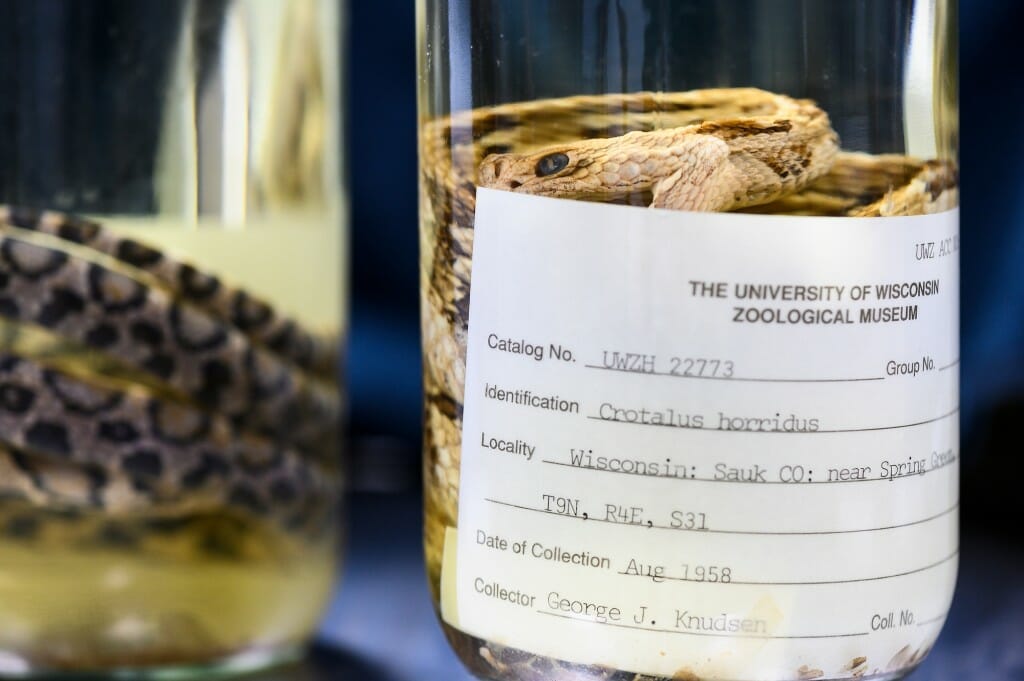
In 2008, scientists discovered a fungus that was killing endangered snakes in Illinois, but preserved snakes in the University of Wisconsin Zoological Museum show that the fungus was already widespread in the United States more than 75 years ago.
Jeff Lorch, a microbiologist at the U.S. Geological Survey’s National Wildlife Health Center in Madison, looked for the fungus, Ophidiomyces ophidiicola, on museum specimens collected decades before the fungus was isolated from Eastern massasauga rattlesnakes being studied for (eventually successful) inclusion as a protected species.
“What we were worried about around 2008 was whether this fungus was just introduced from somewhere else in the world,” says Lorch. “Those are the pathogens that concern you most from a wildlife disease management perspective, because a native species may have no protection against something that comes from far away, that it’s never seen before.”
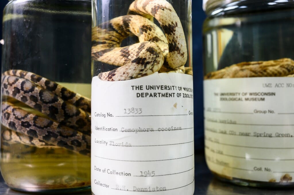
Lorch examined more than 500 snakes in jars, preserved specimens at UW–Madison’s Zoological Museum and the former Morehead State University Museum Collection, looking for telltale lesions on the snakes’ skin. About 9 percent of them showed signs of infection by the potentially deadly fungus.
Lorch took thumbnail-sized skin samples from several, including six from UW–Madison. Three of those — a scarlet snake collected in Florida in 1945, a timber rattlesnake found in Wisconsin in 1958 and a gray rat snake from Tennessee circa 1973 — yielded tissue from which extracted DNA confirmed the presence of the fungus.
“That’s a wide distribution across the eastern U.S. Not just a single snake or place or time, and well before the disease was confirmed,” says Lorch, who published his findings this summer in the journal Emerging Infectious Diseases. “It changes the way we consider the fungus, the direction we may take with research and management, and that’s thanks to museum collections like the one here at UW.”
There are more than 700,000 specimens — from tiny mussel shells to hulking hippo skulls — at the UW Zoological Museum, which makes them available on campus and loans them to institutions and scientists all over the world for teaching and research purposes like Lorch’s study, according to Laura Monahan, the museum’s curator of collections.
“They can’t go back in time to collect samples for their work,” Monahan says. “But we can preserve these snapshots in time. If they’re preserved well, there are so many things researchers can see in them.”
Lorch took samples from as early as 1929 from the Morehead State collection that looked like they had fungal infections, but those snake skins were not preserved in a way that would still give up DNA.
“You can still see the fungus in the layers of skin in the UW specimens. You can still see the cell structure,” he says. “It’s always amazing to me how well preserved these specimens are.”
That’s a tricky part of growing and maintaining museums like Monahan’s.
“The people who collected things we have from the 1800s didn’t know how we would use them. We don’t really know how they might be used in the future,” she says. “But we can be confident they will continue to figure in new science even as they age.”
news.wisc.edu/uw-snake-specimens-show-deadly-fungus-has-been-around-for-decades/
August 23, 2021 By Chris Barncard For news media
news.wisc.edu/content/uploads/2021/08/Zoology_snakes21_4540-1024x681.jpgIn 2008, scientists discovered a fungus that was killing endangered snakes in Illinois, but preserved snakes in the University of Wisconsin Zoological Museum show that the fungus was already widespread in the United States more than 75 years ago.
Jeff Lorch, a microbiologist at the U.S. Geological Survey’s National Wildlife Health Center in Madison, looked for the fungus, Ophidiomyces ophidiicola, on museum specimens collected decades before the fungus was isolated from Eastern massasauga rattlesnakes being studied for (eventually successful) inclusion as a protected species.
“What we were worried about around 2008 was whether this fungus was just introduced from somewhere else in the world,” says Lorch. “Those are the pathogens that concern you most from a wildlife disease management perspective, because a native species may have no protection against something that comes from far away, that it’s never seen before.”
Lorch examined more than 500 snakes in jars, preserved specimens at UW–Madison’s Zoological Museum and the former Morehead State University Museum Collection, looking for telltale lesions on the snakes’ skin. About 9 percent of them showed signs of infection by the potentially deadly fungus.
Lorch took thumbnail-sized skin samples from several, including six from UW–Madison. Three of those — a scarlet snake collected in Florida in 1945, a timber rattlesnake found in Wisconsin in 1958 and a gray rat snake from Tennessee circa 1973 — yielded tissue from which extracted DNA confirmed the presence of the fungus.
“That’s a wide distribution across the eastern U.S. Not just a single snake or place or time, and well before the disease was confirmed,” says Lorch, who published his findings this summer in the journal Emerging Infectious Diseases. “It changes the way we consider the fungus, the direction we may take with research and management, and that’s thanks to museum collections like the one here at UW.”
There are more than 700,000 specimens — from tiny mussel shells to hulking hippo skulls — at the UW Zoological Museum, which makes them available on campus and loans them to institutions and scientists all over the world for teaching and research purposes like Lorch’s study, according to Laura Monahan, the museum’s curator of collections.
“They can’t go back in time to collect samples for their work,” Monahan says. “But we can preserve these snapshots in time. If they’re preserved well, there are so many things researchers can see in them.”
Lorch took samples from as early as 1929 from the Morehead State collection that looked like they had fungal infections, but those snake skins were not preserved in a way that would still give up DNA.
“You can still see the fungus in the layers of skin in the UW specimens. You can still see the cell structure,” he says. “It’s always amazing to me how well preserved these specimens are.”
That’s a tricky part of growing and maintaining museums like Monahan’s.
“The people who collected things we have from the 1800s didn’t know how we would use them. We don’t really know how they might be used in the future,” she says. “But we can be confident they will continue to figure in new science even as they age.”